Cancer treatment has advanced significantly over the years, offering patients new options that are less invasive and highly effective. One such innovation is microwave ablation, a targeted treatment used to destroy cancerous lesions in organs such as the liver without the need for major surgery.
Liver metastasis, which occurs when cancer spreads from another organ to the liver, is a common challenge in oncology. While traditional treatments like surgery and chemotherapy remain important, modern image-guided procedures now allow specialists to treat selected lesions with precision and minimal recovery time.
This case study highlights how a patient with liver metastasis successfully underwent microwave ablation at Dev Hospital of Vascular and Interventional Radiology, demonstrating how advanced interventional radiology techniques can help manage complex cancer conditions.
Understanding Liver Metastasis
Liver metastasis occurs when cancer cells from another part of the body travel through the bloodstream or lymphatic system and form tumours in the liver. The liver is one of the most common sites for metastatic cancer due to its rich blood supply.
Among the cancers that frequently spread to the liver is colorectal cancer, particularly cancers originating in the sigmoid colon.
Patients with liver metastasis may experience symptoms such as:
- Abdominal discomfort or pain
- Fatigue or weakness
- Unexplained weight loss
- Loss of appetite
- Abnormal liver function tests
In many cases, however, liver metastases may remain asymptomatic and are detected during routine imaging scans performed during cancer follow-up.
Early detection plays an important role in determining the most appropriate treatment strategy.
Patient Background: A Recurrence After Initial Cancer Treatment
A 61-year-old female patient with a previous history of carcinoma of the sigmoid colon presented for evaluation.
Approximately one year earlier, the patient had undergone surgical resection of the primary colon tumor along with treatment for liver metastasis. Following surgery, she completed a full course of chemotherapy as part of her cancer management plan.
During routine follow-up evaluation, imaging revealed a new solitary lesion in segment VIII of the liver, indicating a recurrence in the form of liver metastasis. Since the lesion was isolated and localized, the medical team evaluated options for targeted minimally invasive treatment.
Investigating the Liver Lesion
To assess the nature and extent of the lesion, the patient underwent a Positron Emission Tomography (PET) scan, which is commonly used in oncology to detect metabolically active cancer cells.
The PET scan findings confirmed:
- A solitary metastatic lesion in segment VIII of the liver
- No evidence of widespread metastatic disease elsewhere
A Modern Treatment Option: Microwave Ablation
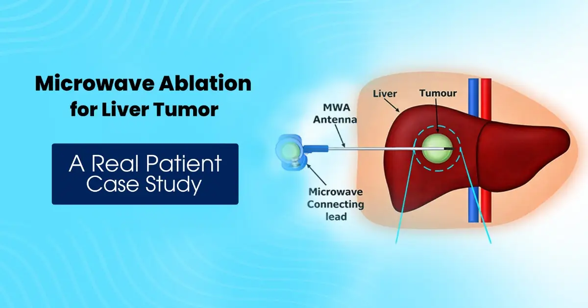
After reviewing the imaging findings and medical history, the interventional radiology team recommended microwave ablation, a minimally invasive procedure designed to destroy tumors using heat energy.
Microwave ablation works by inserting a thin probe directly into the tumor under imaging guidance. The probe emits microwave energy, generating heat that destroys cancer cells while preserving the surrounding healthy tissue.
Advantages of Microwave Ablation
Compared to traditional surgery, microwave ablation offers several benefits:
- Minimally invasive procedure
- Precise targeting of the tumor
- Shorter hospital stay
- Reduced recovery time
- Lower risk of complications
For selected patients with small or isolated liver metastases, this treatment can provide excellent results.
The Procedure: Image-Guided Precision Treatment
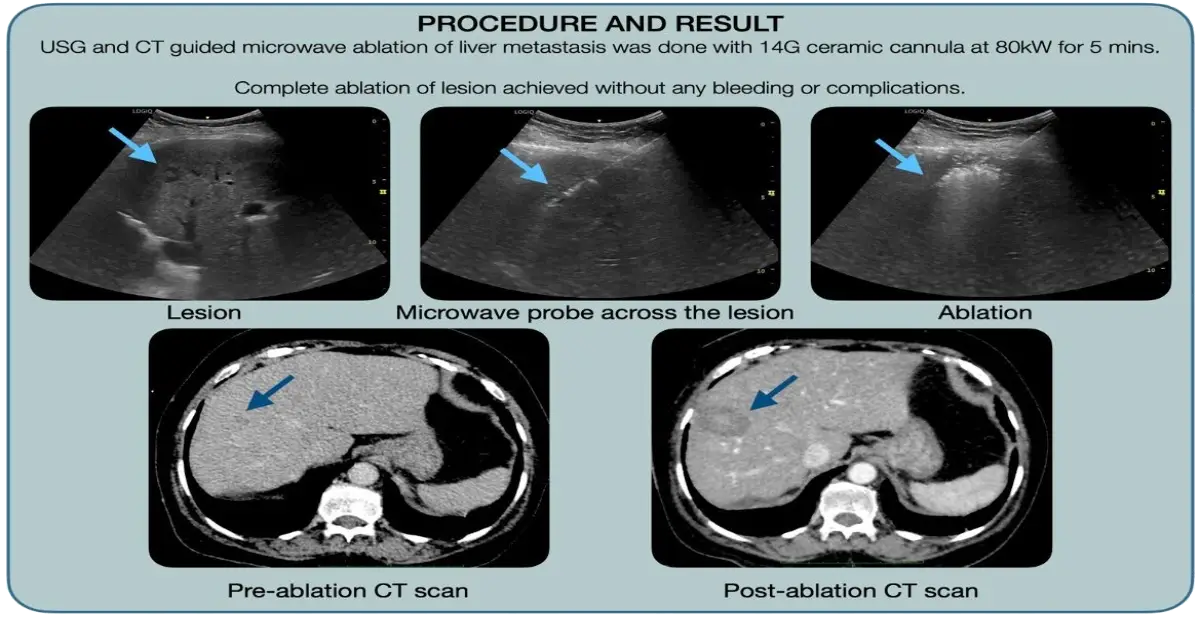
The procedure was performed using ultrasound (USG) and CT guidance, ensuring accurate placement of the ablation probe within the tumor.
The key steps included:
- A 14G ceramic microwave ablation cannula was carefully inserted into the liver lesion.
- Microwave energy was delivered at 80 kW for approximately 5 minutes.
- The generated heat destroyed the tumor cells within the targeted region.
- Then track ablation was done at 40 kW and cannula was removed.
The procedure was completed successfully, and complete ablation of the lesion was achieved. Procedure was performed under General Anesthesia; post procedure patient was awaken. Importantly, there were no bleeding events or procedural complications, demonstrating the safety of this image-guided technique.
Post-Procedure Imaging
Imaging plays an important role in accessing effectiveness of the procedure. Immediate post procedure CT scan is done to see for precise treatment delivery and to see for any complications including bleeding.
Follow up CT scan is done at 1, 3, 6 months and then every 6 monthly.
Recovery and Outcome
Because microwave ablation Treatment is a minimally invasive procedure, recovery is generally quicker compared to traditional surgery.
In this case:
- The procedure was completed without complications
- The patient tolerated the treatment well
- Imaging confirmed complete tumor ablation
Such outcomes demonstrate the effectiveness of image-guided cancer treatments in selected patients.
The Role of Interventional Radiology in Cancer Care
Interventional radiology is transforming the way many diseases are treated, including cancer.
Using advanced imaging technologies such as ultrasound, CT scans, and fluoroscopy, specialists can perform highly precise procedures that treat tumors with minimal disruption to the body.
These techniques are particularly valuable for:
- Liver tumors and cancer
- Kidney tumors and cancer
- Lung cancer
- Head and neck tumor and cancers
By offering targeted treatment options, interventional radiology helps reduce recovery time while maintaining high treatment success rates.
Expert Care at Dev Hospital of Vascular and Interventional Radiology
Prompt referral to a specialized center with an experienced interventional radiologist Ahmedabad ensures optimal planning and execution. Advanced minimally invasive procedures require experienced specialists and modern imaging facilities.
At Dev Hospital of Vascular and Interventional Radiology, patients receive treatment from highly trained experts, including:
Dr. Milan Jolapara (MD, DM)
A specialist in vascular and interventional radiology with expertise in advanced minimally invasive procedures, including tumor ablation therapies.
Dr. Trupti Mehta (MD, FVIR)
An experienced interventional radiologist skilled in image-guided procedures used for treating vascular disorders and cancer-related conditions.
When Should Liver Metastasis Be Evaluated by Specialists?
Patients with a history of cancer should undergo regular follow-up imaging to monitor for recurrence or metastasis.
You should consider consulting a specialist if:
- You have a history of colorectal cancer or other cancers
- Follow-up scans detect suspicious liver lesions
- You are exploring minimally invasive treatment options for tumours
- You want a second opinion regarding advanced cancer treatments
Early detection and timely intervention can significantly improve treatment outcomes. To explore the complete range of services, visit Our Treatments.
Consult Our Specialists
If you or a loved one has been diagnosed with liver metastasis or requires advanced image-guided treatment, expert consultation can help determine the most appropriate treatment plan. The team at Dev Hospital of Vascular and Interventional Radiology offers modern minimally invasive procedures designed to treat complex conditions safely and effectively. Consulting with experienced specialists can help you understand your options and receive personalized care based on your medical needs.