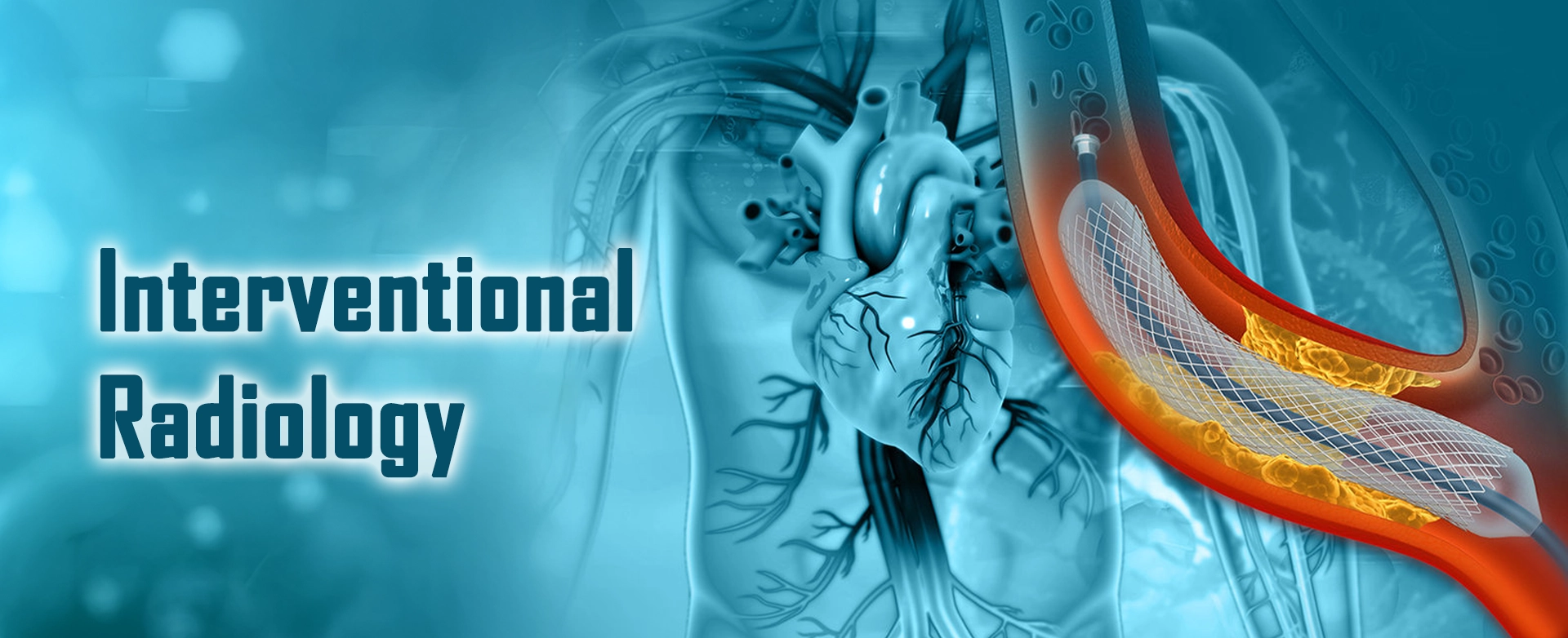
Interventional Radiology utilizes minimally-invasive image-guided technique to diagnose and treat nearly every organ system. With the enhancement of technology, the interventional radiology procedures are now a boon to modern medicine.
The objective of Interventional radiology is to minimize risk to the patient and improve health outcomes. Interventional Radiologists can deliver treatments for various-cancers, fibroids, back and joint pain, varicose veins, peripheral arterial diseases, kidney and bile duct disease to name a few.
Interventional Radiologists are image-guided therapy clinicians who are specially trained in image interpretation and minimally invasive treatments of a wide variety of conditions across multiple specialties.
What procedures do interventional radiologists perform?
Interventional radiologists do a variety of procedures, including:
- Angiography: This is an X-ray of the arteries and veins to find blockage or narrowing of the vessels, as well as other problems.
- Angioplasty: The doctor puts a small balloon-tipped catheter into a blood vessel. Then he or she inflates the balloon to open up an area of blockage inside the vessel.
- Embolization: The doctor puts a substance through a catheter into a blood vessel to stop abnormal blood flow or leakage through that vessel. This can be done to control bleeding. A Catheter-based technique using coils, particles or glue to block tumour vessels or acute bleeding is called embolisation. In case of tumour embolisation, viability of a tumour is dependent on its blood supply. With embolisation, we can instill intra-arterial chemotherapy into the tumour and then block the blood supply of the tumour by embolising its blood vessels. The tumour gradually shrinks in size and dies off.
- Chemoembolization: Same as Embolisation but using local targeted high dose chemotherapy to treat tumours.
- Ablation: Needle or catheter-based technique using thermal energy forms like radiofrequency, microwave or cryo technique to treat tumours or varicose veins.
- Gastrostomy tubes: The doctor puts a feeding tube into the stomach if you can’t take food by mouth.
- Intravascular ultrasound: The doctor uses ultrasound to see inside a blood vessel to find problems.
- Stent placement: The doctor places a tiny mesh coil (stent) inside a blood vessel at the site of a blockage. He or she expands the stent to open up the blockage.
- Balloons and stents: Techniques to open up a blocked tube like an artery, vein, bile duct, ureter, colon, or esophagus.
- Foreign body removal: The doctor puts a catheter into a blood vessel to remove a foreign body from the vessel.
- Needle biopsy: The doctor puts a small needle into almost any part of the body, guided by imaging techniques like ultrasound or CT scan, to take a tissue biopsy. This type of biopsy can give a diagnosis without surgery. An example of this procedure is called the needle breast biopsy.
- IVC filters: The doctor puts a small filter into the inferior vena cava (IVC). This is a large vein in your abdomen. The filter catches blood clots that may go into your lungs.
- Injection of clot-dissolving medicines: Acute thrombosis within any artery or vein of the body can be a life-threatening condition. The doctor injects clot-dissolving medicines such as tissue plasminogen activator. This procedure is called Thrombolysis. This medicine dissolves blood clots and increases blood flow to your arms, legs, or organs in your body.
- Catheter insertions: The doctor puts a catheter into a large vein to give chemotherapy medicines, nutrition, or hemodialysis. He or she may also put in a catheter before a bone-marrow transplant.
Advantages
- Day care or short admission procedures
- Less / no pain
- More cosmetic – pinhole/spot dressing surgery
- Less morbidity/complications
- More rapid return to work
- Does not close the door for conventional surgeries
- Can be combined with other treatments like chemotherapy
Conditions that can be treated include
- Uterine fibroids
- Rest pain and claudication in case of Ischaemic diabetic foot
- More cosmetic – pinhole/spot dressing surgery
- Varicose veins
- Male varicocele and pelvic congestion syndrome
- Benign prostatic hyperplasia – Prostatic enlargement in elderly
- Recurrent haemoptysis, GI bleeds
- Back pain
- Curative treatment in form of ablation for small kidney, lung or liver-procedures tumours
- Local control for larger tumours
- Venous access and ports
- Renal dialysis access
- Bile duct and ureteric blocks – Tube insertion or stenting
- Management of liver-procedures cirrhosis
What are the benefits of interventional radiology?
As interventional radiology is minimally invasive, patients experience less
pain. The incisions that may be made are quite small and won’t need stitches or
large bandages to heal. The risk of infection or other complications is quite
low in interventional radiology procedures. The treatment can be precisely
guided to the diseased organs, arteries and veins without causing any damage to
the surrounding organs. The recovery time from an interventional radiology
procedure is quite short as well.
Will I be awake during my interventional radiology procedure?
Yes, you will probably be awake during the interventional radiology procedure.
Unlike traditional surgery, patients aren’t given general anaesthesia during the
procedure. What will instead happen is your care team will numb the area of the
incision with a local anaesthetic to eliminate any discomfort. They will then
use an intravenous line to deliver sedation to make sure you are relaxed and
comfortable during the procedure.